稳定性—— 为长期成像实验提供可靠的性能。
生命科学
物理科学
 牛津仪器集团成员

由Oxford Instruments开发的Fusion,开启了共聚焦显微镜成像软件的新纪元。Fusion专为与我们的高性能硬件完美配合而设计,为控制共聚焦成像系统(包括Benchtop-BC43和Dragonfly)以及光刺激设备(如Mosaic)提供了一个统一且高度灵活的平台。
Request Trial License Upgrade Fusion
Fusion 的设计宗旨是优先考虑用户需求、灵活性和简便性。它使台式显微镜和 Dragonfly 系统都能在极少培训的情况下轻松操作,从而高效地执行复杂的多维实验。
Fusion 将图像采集、可视化和数据处理整合于一个直观的平台中,从而简化成像流程。其引导式工作流和自适应界面可满足各层次用户的需求,帮助用户快速从设置阶段过渡到获得高质量结果。凭借智能自动化和响应迅速的性能,Fusion 在最大限度缩短设置时间的同时,确保输出结果的一致性。
稳定性—— 为长期成像实验提供可靠的性能。
易用性——学习曲线平缓,用户能快速上手。
灵活性——支持多种应用,例如多维实验、刺激后测试等
图像分析——为了实现高级处理、分析工作流和数据定量分析 ,Fusion Software 与 Imaris 实现了无缝集成 。
模块化——可根据用户需求调整Software,并随需求变化进行升级。
超分辨率——兼容超分辨率协议,包括 SMLM 和 SRRF Stream+。
硬件集成—— BC43台式共聚焦显微镜、Dragonfly和Mosaic均 通过Fusion Software进行操作。
图像处理—— 集成了采集过程中所需的多种基本图像处理工具,例如拼接和跨序列分析。
Fusion 模块专为简单和复杂的协议而设计,致力于提升工作效率并确保易用性。请查看每个模块,了解其功能如何提升您的图像采集效果。
点击“+”号,了解更多关于 Fusion 功能的信息。
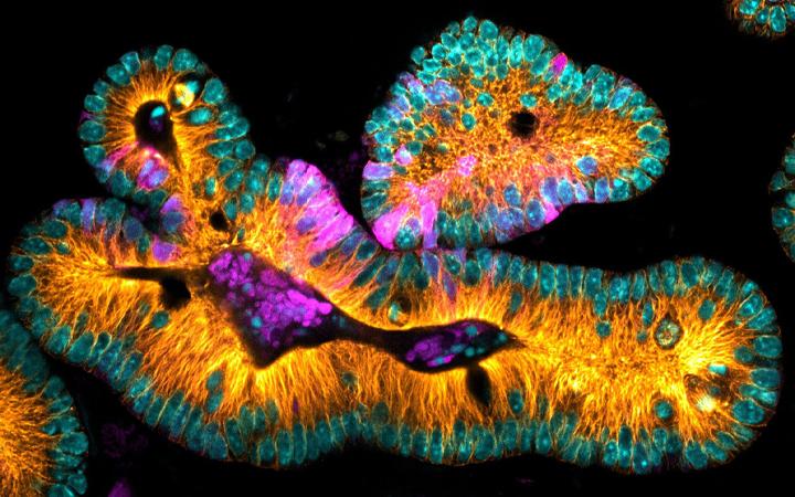
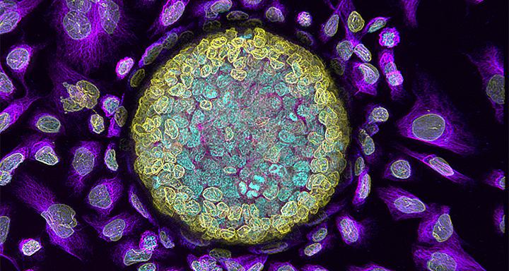
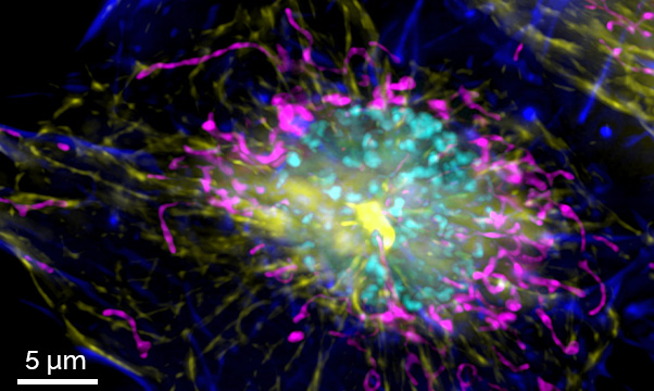
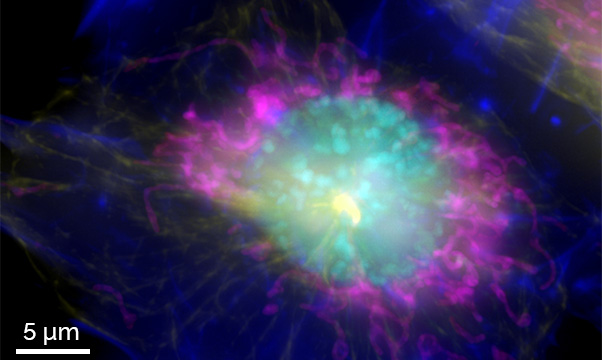
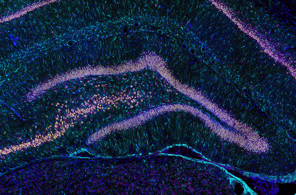
Image Legend: Mouse Intestinal Organoid expressing LGR5-GFP to visualize stem cells (magenta) microtubules (orange) and DNA (cyan). Image courtesy of Nicole Dawney from the Bergstralh Lab and High Content Imaging Core, University of Rochester.
Deeper Insight, Better Data
*1 Piezo available in Dragonfly Fusion only
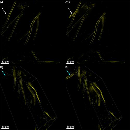
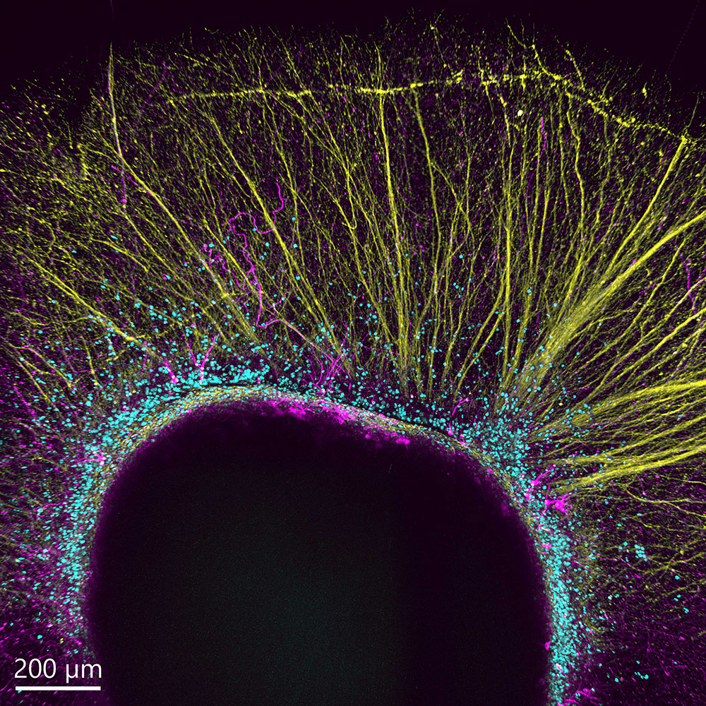
Image Legend – The Deep Z feature in Fusion reveals hidden structures at depth
A Z‑stack was acquired (A, B), and the Deep Z feature was applied (A1, B1). In panels A and B, the laser power was kept constant at 6%. In panels A1 and B1, the laser power started at 6%and increased exponentially up to 30%. Deep Z activation occurred at approximately 80 µm depth, after which the laser power ramped progressively to 30%.
A total of 231 stacks were acquired, corresponding to a 137‑µm imaging range. Structures that become visible due to the increasing laser power are indicated by the white arrows in A), A1)(MIP) and by the blue arrows B), B1) (orthogonal view).
Immediate experimental feedback
Grasp the dynamics of biology
Translate cell behaviours into biological insights by imaging dynamic processes in living cells.
*Available in Dragonfly only
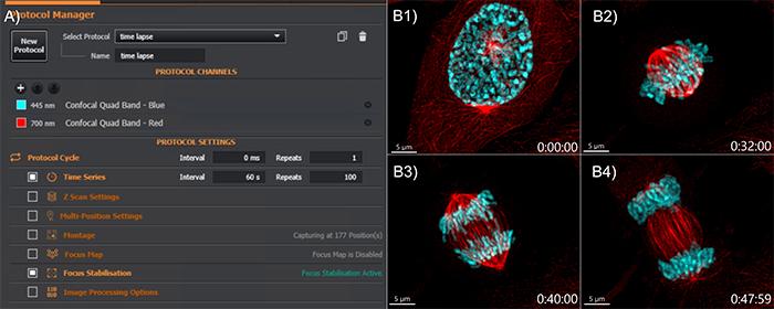
Figure Legend – Time-lapse setup and results
(A) Easy time‑lapse setup: select the time interval and number of repeats. Focus stabilization can be activated for higher positional accuracy. The quad‑band filter enable seven faster image acquisition. (B) Mammalian cell division sequence: (B1) Prophase, (B2)Metaphase, (B3) Anaphase, (B4) Telophase. Microtubules are shown in red, and DNA in cyan.
Stay sharp. Stay confident. Stay in focus.
Fusion control hardware-based autofocus maintains sample focus throughout the experiment. This feature is critical for live imaging assays, multi-well plate imaging, large fixed samples, multi-position imaging, and multi-tile imaging.
Reliable. Accurate. Always in focus
Multi-position Imaging of Mammalian Cells with BC43.
At each time point, four positions were captured in parallel, each with three channels and 15 Z‑stacks. Shown side‑by‑side, the video highlights how the Focus Seek & Lock technology keeps every position perfectly in focus—even during rapid multi‑site navigation. Image credits: Ines Baião‑Santos, Álvaro Tavares (Universidade do Algarve); Claudia Florindo (Oxford Instruments).
Expand your imaging capabilities
Configure multiple independent acquisition experiments and automate acquisition control for external devices.
Note – This is an advanced feature, and its use requires an understanding of Python coding
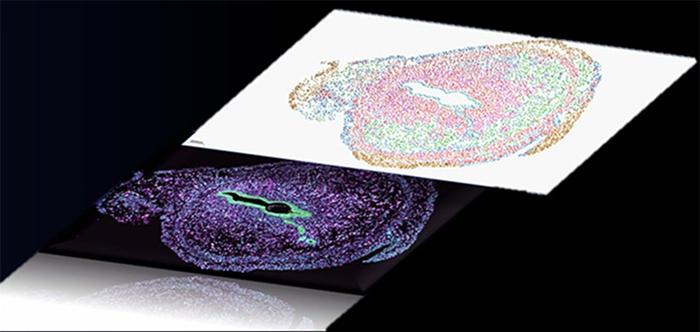
Spatial omics graphic representation
With the power of our REST API, users can orchestrate complex workflows by linking multiple protocols and external devices. Comprehensive gene-expression atlases can be generated by coordinating our spinning-disk confocal systems (Dragonfly or BC43) with automated microfluidic triggering.
Image Legend: Mouse Intestinal Organoid expressing LGR5-GFP to visualize stem cells (magenta) microtubules (orange) and DNA (cyan). Image courtesy of Nicole Dawney from the Bergstralh Lab and High Content Imaging Core, University of Rochester.
Effortless Sample Overview
See your entire sample instantly. With one click, our smart spiral montage delivers a complete, high-quality overview—no missed areas, no duplicate tiles.
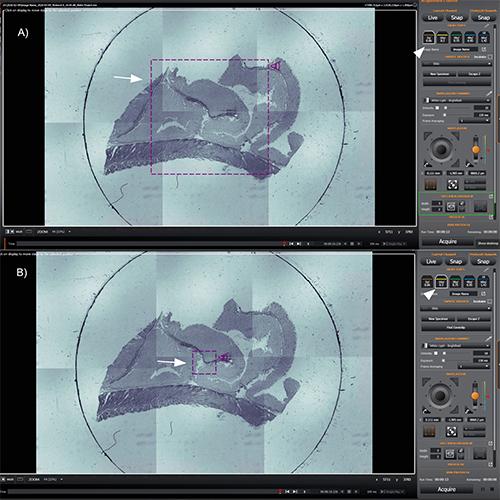
Image Legend – Overview of sample navigation & spiral montage
A) A 2×3 spiral montage was acquired using the 2× objective. The navigation icon (arrow), the 2× objective selection (arrowhead), and the montage definition (green rectangle) can be observed. B) When switching to the 10×objective (arrowhead), the field of view is automatically adjusted (arrow).
See Beyond the Field of View
Unlock the complete perspective of your sample with seamless, high‑coverage imaging.
Irregular montage new - Imaging That Adapts to Your Sample
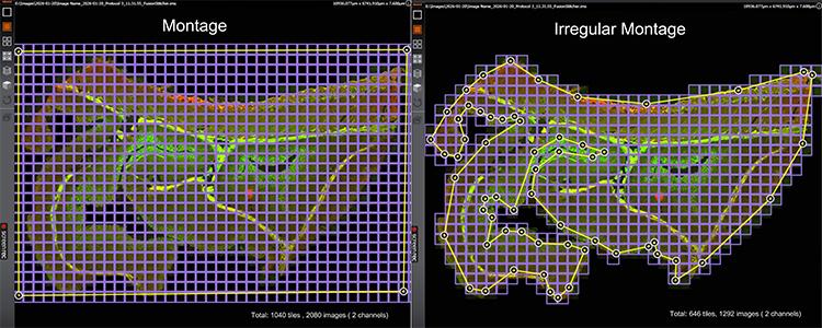
Figure Legend – Irregular montage increases productivity
This image compares a standard montage (left) with an irregular montage on theBC43 system (right). The standard montage requires 1040 tiles (2080 images, 2channels). This strategy allows imaging well beyond the field of view, enabling the capture of the entire sample at the desired resolution.
The irregular montage needs only 646 tiles (1292 images). This results in a two‑fold productivity increase for 2D imaging, with even greater gains when acquiring z‑stacks or additional channels
Increase Productivity Through Higher Sample Throughput
Boost the efficiency of every experiment and extract more insight from each run.
1. More Output per Experiment - Deliver greater insight from every run without increasing workload.
2. Focus on What Matters - Acquire only the most relevant cells or sample regions to maximise efficiency.
3. Scale Further with Montage - Combine targeted acquisition with montage imaging for even higher coverage and context.
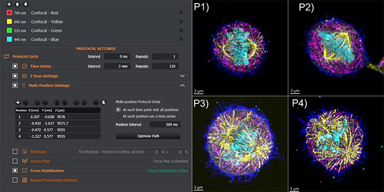
Multiple positions increase experimental throughput
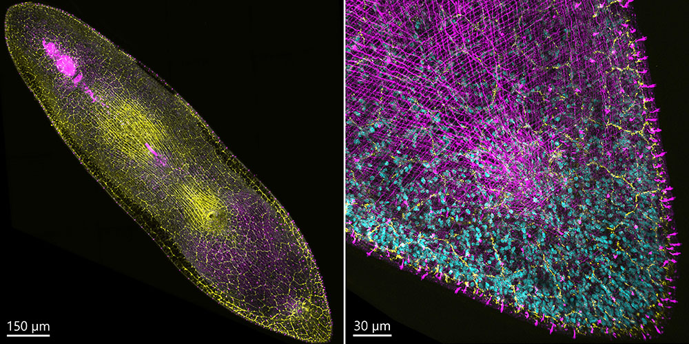
In this time‑lapse, multichannel experiment, four positions were acquired to track mitotic cells, which can be rare depending on the tissue and cell line. The protocol included four confocal channels, time‑lapse, Z‑stack, and multi‑position acquisition with active focus stabilisation. Montage acquisition can also be added to further boost throughput.P1–P4 (right panels) show the four mitotic cells. Colour coding: dark blue – actin, pink – mitochondria, yellow – microtubules, cyan – DNA.
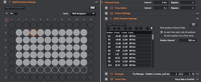
Redefine Throughput in Multi-Well Assays
Efficiently image and analyse multiple experimental conditions, reducing turnaround time and increasing throughput.
One click well selection
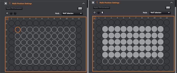
Navigate with well plate view or table view
All protocol combinations are possible.
Keep every field in focus
Enables correction of tilt and sample unevenness. Can be used in multi-position protocols.
Supports robust acquisition of uneven and complex samples.
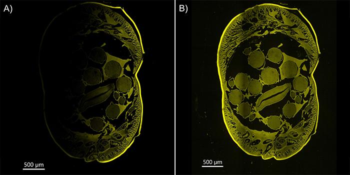
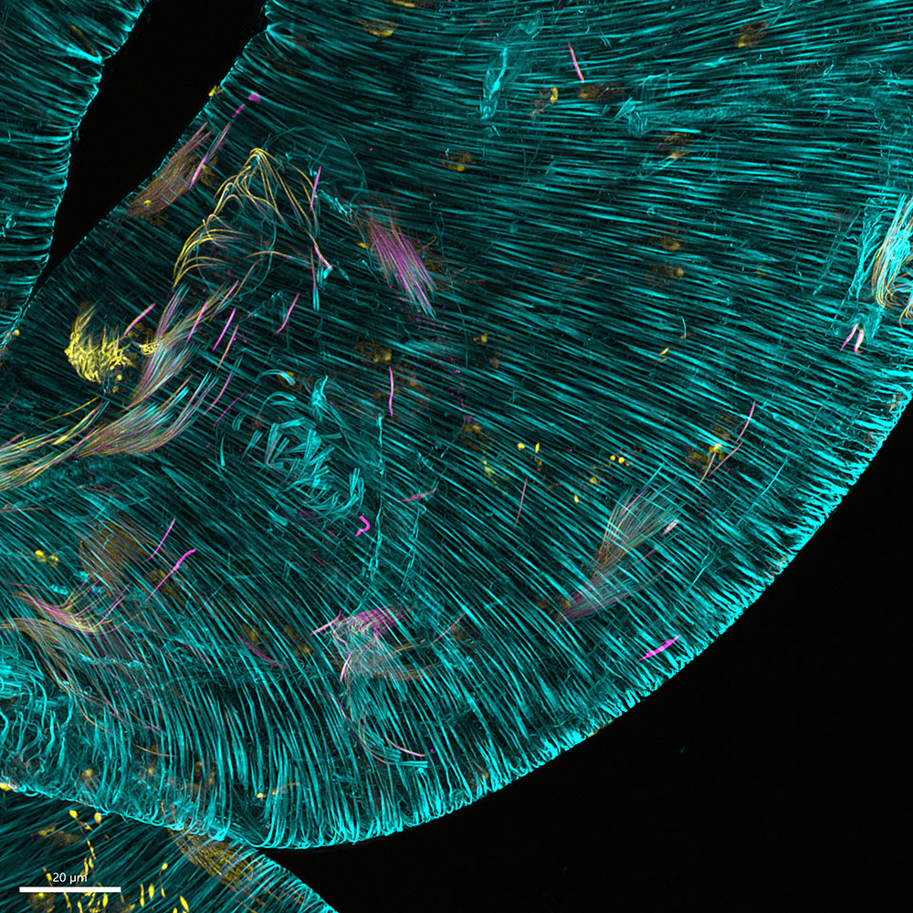
Figure Legend – Focus Map Correction of Slide Tilt and Sample Unevenness
Twelve tiles of the Ascaris sample were acquired. (A) Due to tilt in the slide, the left side of the image is not well focused and appears less visible. (B) After applying the Fusion Focus Map to the tilted Ascaris sample, both the left and right regions of the specimen appear sharply in focus.
Image Legend: Mouse Intestinal Organoid expressing LGR5-GFP to visualize stem cells (magenta) microtubules (orange) and DNA (cyan). Image courtesy of Nicole Dawney from the Bergstralh Lab and High Content Imaging Core, University of Rochester.
Illuminate Function, Frame by Frame
An integrated, precise, and flexible photostimulation workflow for advanced functional imaging.
Compatibility: Dragonfly + Mosaic systems.
Perform Precise Photostimulation
Take precise control of photostimulation delivery. Target areas of tissues, individual cells or intracellular features. Gain insights into processes within living cells with powerful techniques such as photoactivation, FRAP, uncaging and optogenetics.
Simple, and accurate B-TIRF control.
B‑TIRF (Borealis TIRF) brings precision TIRF imaging to every user by simplifying a traditionally complex technique. With effortless setup and smart control, it delivers consistent, high-contrast and uniform illumination for demanding single-molecule and membrane-level studies.
SMLM or SRFF-Stream
Single‑molecule localization microscopy (SMLM) or push‑button super‑resolution—whichever fits your workflow.
SMLM – Single‑Molecule Localization Microscopy
(SMLM available for dragonfly only)
SRRF‑Stream – One‑Click Super‑Resolution
Achieves resolutions down to 140 nm
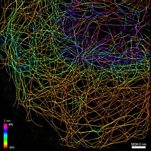
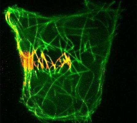
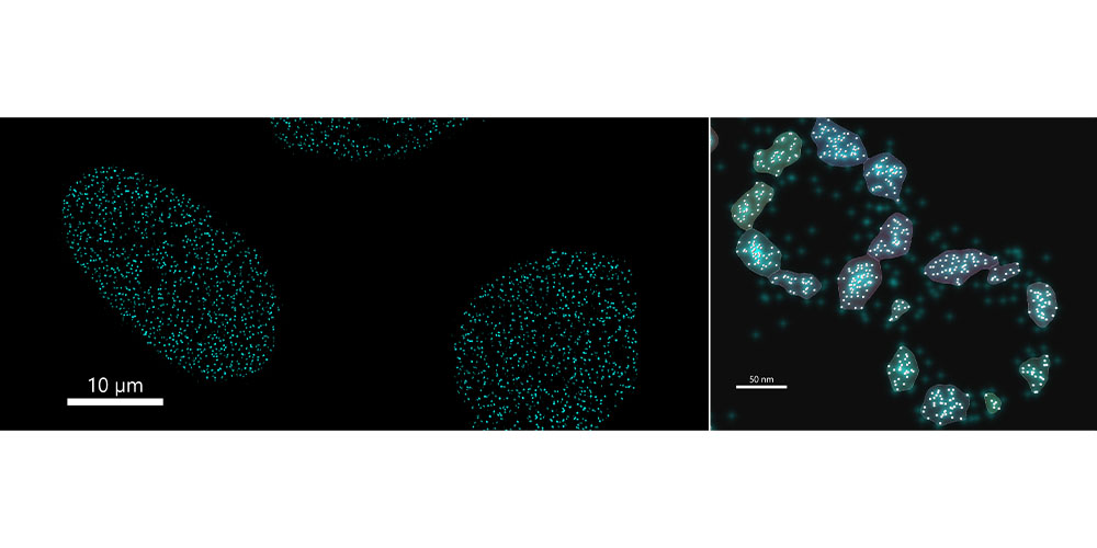
Microtubules in a cell imaged using Single Molecule Localization Microscopy
A 10,000 time‑point series was acquired to capture the stochastic emission of Cy3 fluorophores. The data were rendered, drift‑corrected, and subsequently localized, achieving a median localization precision of ~13 nm. Image acquired with the Andor Dragonfly spinning disk confocal. Data courtesy of Felix Rivera‑Molina, Yale University.
Image Legend: Mouse Intestinal Organoid expressing LGR5-GFP to visualize stem cells (magenta) microtubules (orange) and DNA (cyan). Image courtesy of Nicole Dawney from the Bergstralh Lab and High Content Imaging Core, University of Rochester.
See the big picture by visualising the cells within the context.
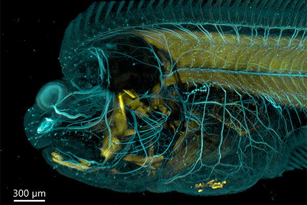
Figure – 3D stitching provides a full overview of the sample
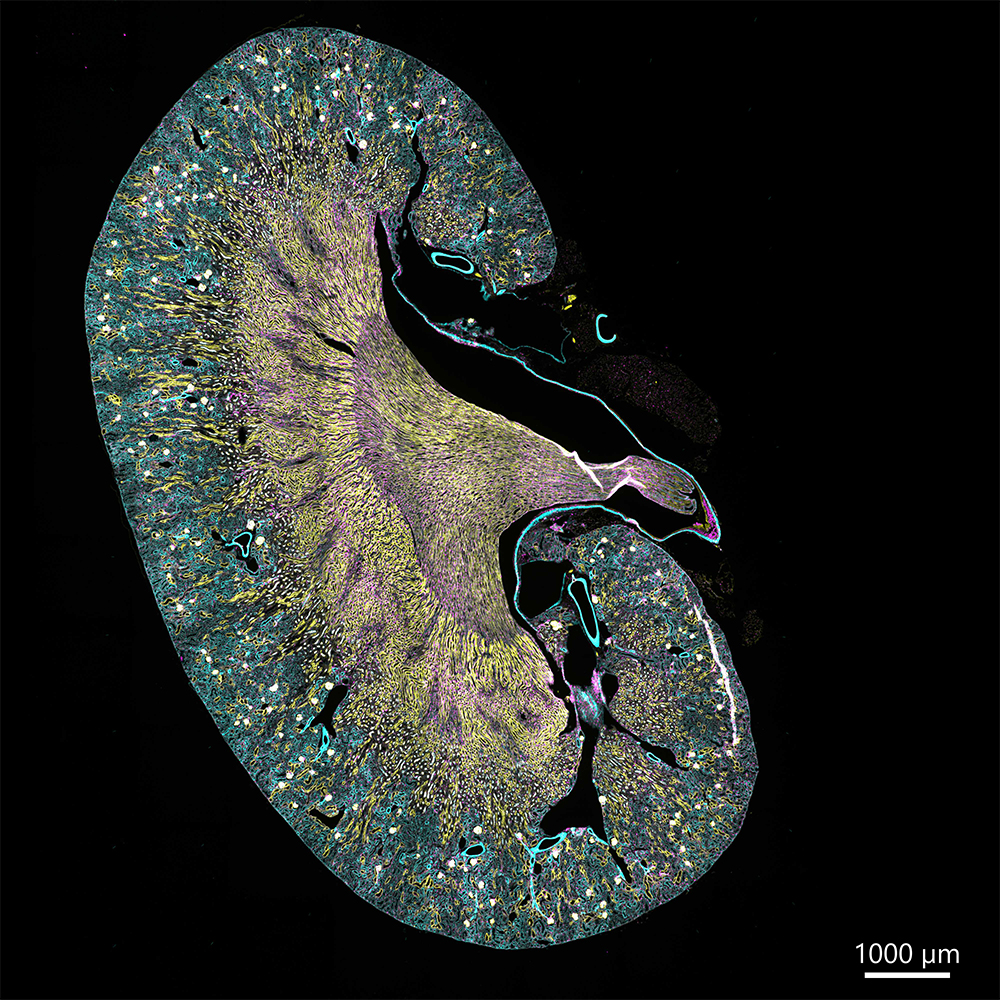
Flat fish imaged on the BC43 using a montage. Six tiles were acquired to compose the image, covering a Z‑range of 554 µm. The 3D image was stitched using the Fusion option activated in the protocol.
(Image credits: Marco Campinho, Universidade do Algarve; Claudia Florindo, Andor Technology)
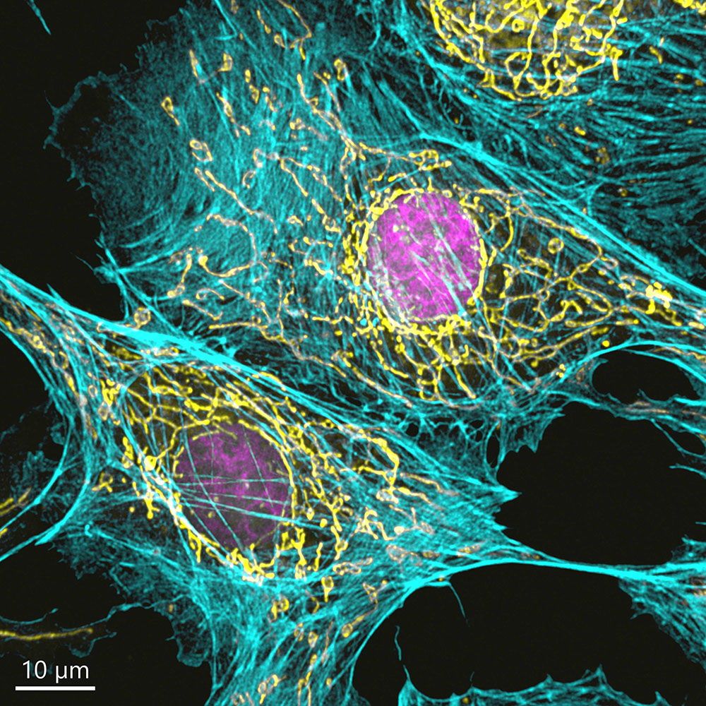
Sharper images, more detail, improved resolution.
ClearViewTM deconvolution enhances image clarity by removing haze and improving resolution.
Enhanced resolution and contrast with ClearView™ deconvolution.
Widefield images of a mammalian cell acquired on the BC43 system demonstrate the improvement in contrast and spatial resolution achieved through ClearView™ deconvolution. Left: Widefield image , right: deconvolved image. Deconvolution was activated in the protocol. Fluorescent labels: actin (dark blue), microtubules (yellow), mitochondria (pink), and DNA (cyan). Image credit: Claudia Florindo, Oxford Instruments.
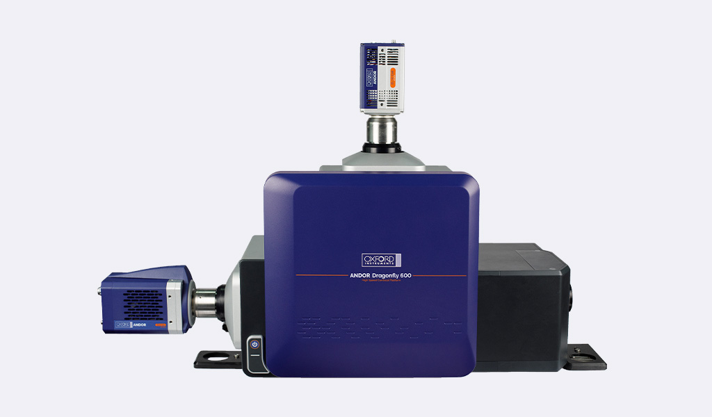
Dragonfly 是一款高速共聚焦平台,能够以卓越的速度和灵敏度,从亚细胞结构到整个生物体,呈现出非凡的图像。它通过单一且功能多样的系统,轻松支持活细胞、大型模式生物以及 SMLM 成像。
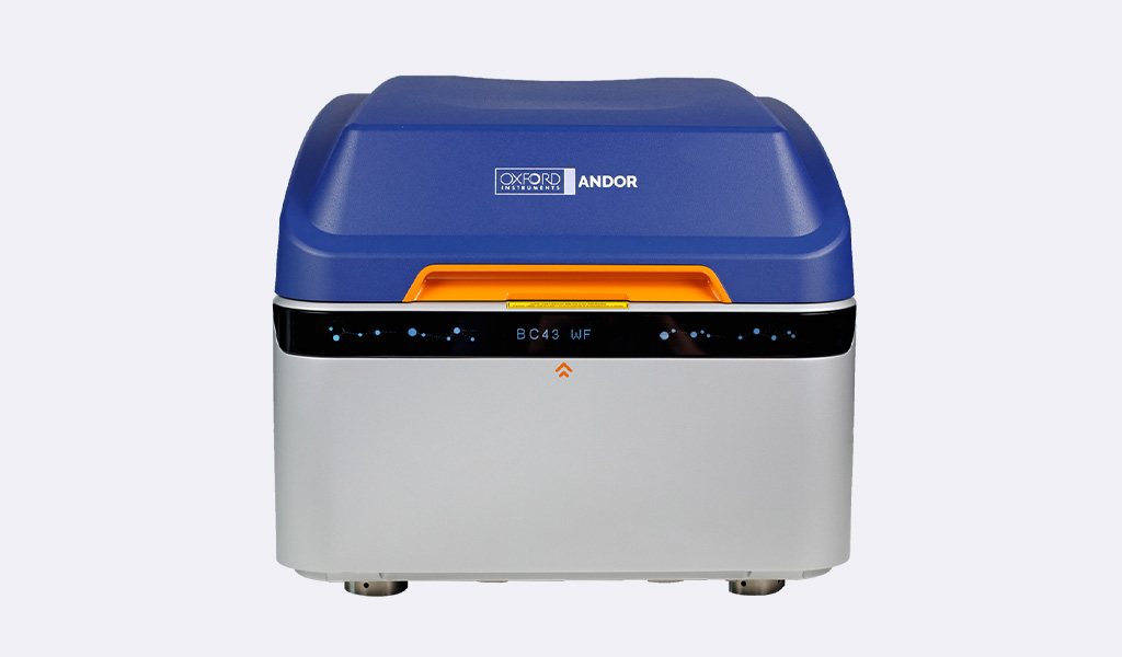
BC43 将入门级的简便性与高端性能融于一体,打造出一款紧凑型台式系统。它支持 2D/3D 宽场成像、共聚焦成像以及一键式超分辨率成像。高速共聚焦成像功能可显著提升工作效率,并由业界领先的认证质量计划提供支持。
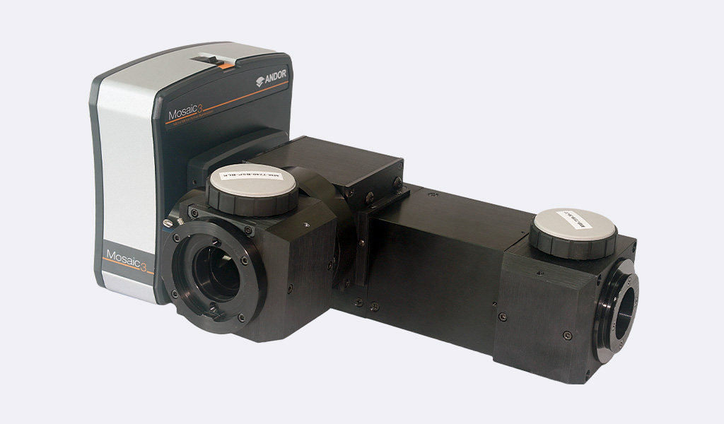
Mosaic 采用数字微镜技术,通过精准、定向的样本照明,助您从研究中获得更多成果。它为强大的光刺激实验开辟了新途径,包括光遗传学、光转换、解笼等众多应用。

© 牛津仪器 2026
 公安机关备案号31010402003473